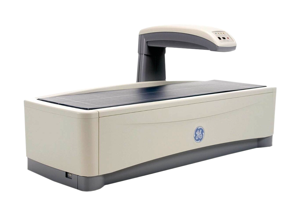
Bone Density
(DEXA)
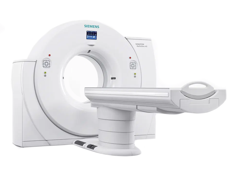
Hi-Speed CT Scan (128 Slice)
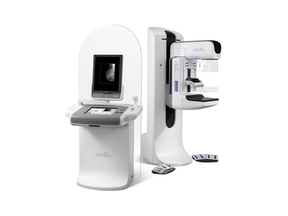
3D Digital Mammography
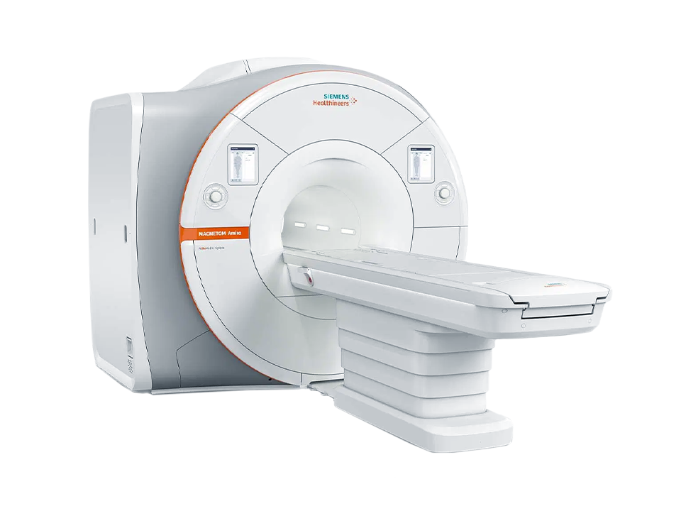
MRI (3T)
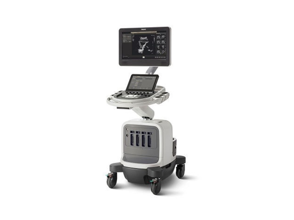
Ultrasound
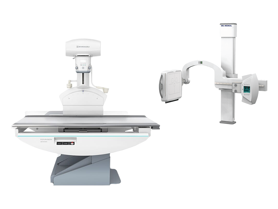
X-Ray/Fluoroscopy


長老計劃


Bone Density (DEXA)

Hi-Speed CT Scan (128 Slice)

3D Digital Mammography

MRI
(3T)

Ultrasound

X-Ray/ Fluoroscopy
長老計劃